
Most people have a clear mental image of an MRI machine: a large white tube that a patient slides into horizontally, lying flat and completely still while the scanner works. That image is accurate for the vast majority of MRI facilities. It is not, however, the only way MRI technology can be configured, and for a significant number of patients, the traditional closed-bore design creates real problems, both in terms of comfort and diagnostic accuracy.
Upright MRI, also called open MRI or stand-up MRI, is a fundamentally different approach to the same technology. Understanding what makes it different, and why that difference matters, helps patients make better-informed decisions about where and how to get their imaging done.
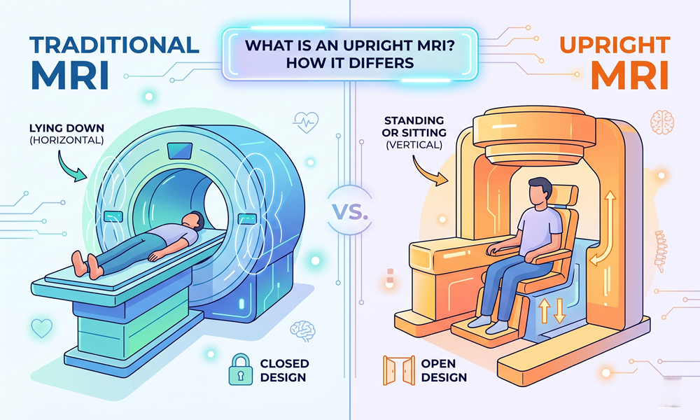
How a Traditional MRI Machine Works
A conventional MRI machine generates a powerful magnetic field and uses radio waves to produce detailed images of the body’s internal structures. The patient lies flat on a motorised table that slides into a narrow cylindrical bore, typically 60 to 70 centimetres in diameter. The bore surrounds the patient on all sides, and the scan takes place while the patient remains as still as possible in that enclosed position.
The closed-bore design produces excellent image quality, and for many types of scans it remains the standard of care. The limitation is that the patient is always scanned in a single position: lying down. For conditions that are positional in nature, meaning symptoms that appear or worsen when the patient is upright, sitting, or bearing weight, that horizontal scan position may not capture the problem at its most visible or clinically significant point.
What Makes Upright MRI Different
An upright MRI machine is designed with an open configuration that allows patients to be scanned while sitting, standing, or in a variety of weight-bearing positions. The magnet surrounds the patient from the sides rather than enclosing them in a tube, which eliminates the claustrophobic environment of a traditional bore scanner. Upright MRI of Deerfield operates one of these systems and is recognised as a leader in open MRI innovation, offering patients an experience that is both more comfortable and, for specific conditions, more diagnostically informative than a conventional scan.
The ability to scan in weight-bearing positions is the most significant clinical advantage. The spine, for example, behaves differently when a patient is upright and gravity is acting on the vertebral column than it does when they are lying flat and decompressed. A disc herniation that is actively compressing a nerve root while the patient is standing may reduce or disappear entirely when they lie down. Scanning only in the supine position risks missing that finding altogether.
Conditions Where Upright MRI Has a Clear Advantage
Spinal stenosis, disc herniations, and spondylolisthesis are among the conditions most likely to present differently depending on patient position. Patients with chronic lower back pain that is consistently worse when upright and relieved when lying down are particularly good candidates for weight-bearing imaging, because the condition producing their symptoms is most visible in the position that triggers them.
Cranio-cervical instability is another area where upright MRI provides diagnostic information that a standard scan cannot. Instability at the junction of the skull and cervical spine is a positional finding by definition. Scanning the patient lying flat, with the head supported and the neck at rest, removes the gravitational load that causes the instability to manifest. An upright or flexion-extension scan captures the anatomy under the conditions that produce the patient’s symptoms.
Beyond spinal conditions, the open configuration is beneficial for larger patients who may not fit comfortably or safely in a standard bore, for elderly patients who find the enclosed environment distressing, and for patients whose anxiety or claustrophobia makes a conventional scan difficult or impossible to complete without sedation.
Image Quality Considerations
A common question from patients and referring physicians is whether upright MRI produces images comparable to those from a closed-bore system. Early open MRI machines used lower field strength magnets that produced images of noticeably reduced quality compared to high-field closed systems. Modern upright MRI technology has narrowed that gap considerably. For musculoskeletal imaging, particularly spinal and joint work, current upright systems produce images of sufficient diagnostic quality for the vast majority of clinical applications.
For certain scan types, particularly brain imaging and some oncological applications, high-field closed systems may still be preferred. For any condition where patient position affects symptom presentation, the clinical value of a weight-bearing scan outweighs field strength considerations, and the upright image showing an active finding is diagnostically more useful than a high-resolution image of anatomy that is not in its symptomatic state.
For patients who have been told they need an MRI, particularly for back, neck, or joint pain that changes with position, understanding why upright MRI is different is a worthwhile step before booking a scan. The technology exists specifically to address the limitations of conventional imaging, and for the right patient and the right condition, it delivers a more complete clinical picture.